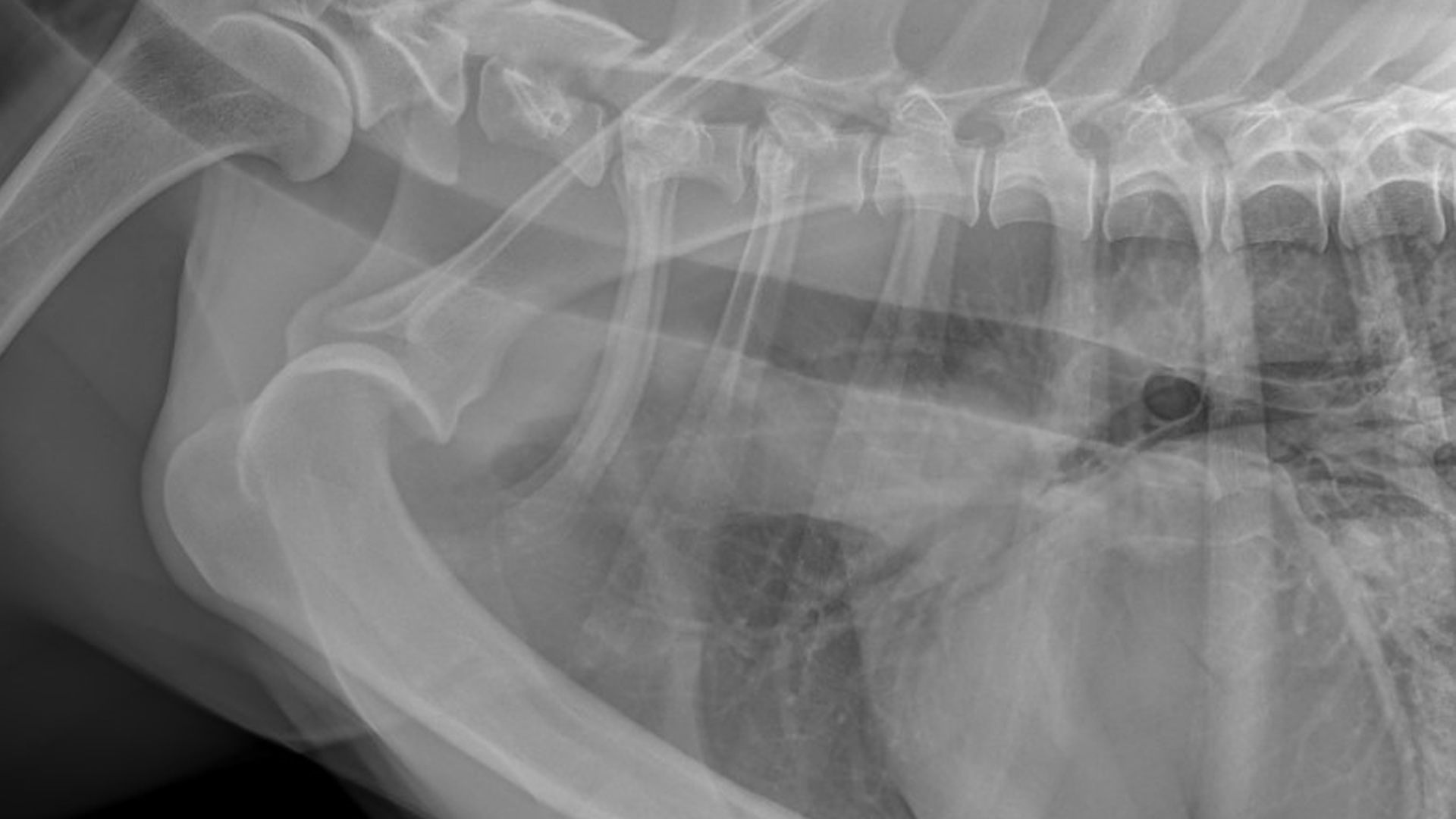
RADIOGRAPH REPORTING
A free service for general
practice vets
Welcome to our radiograph reporting service; we make it our priority to support local practice vets with the management of orthopaedic, neurological and dental cases to help you provide the highest level of care for your patients and clients.
- A surgeon’s interpretation of your radiographs with follow-up advice via phone or email to help you manage the case, including competitive estimates for referral if indicated.
- Please allow five working days for non-urgent advice.
- We always have emergency clinicians available to help with triage and we will do our best to help you as promptly as possible.
Who can use this?
This service is only for veterinary practices with no orthopaedic surgeon, neurologist or dentist on-site. Unfortunately, we are unable to comment on any queries relating to surgery previously performed at another practice, or on cases that are currently under the care of another referral surgeon.
radiograph reporting
How to request
To help us process your radiograph opinion, please follow our guidelines outlined below and email radiographs to:
For files over 50MB in total please send via WeTransfer or Dropbox.
If you believe an urgent opinion is required, please state this in your email subject line and call us on 01483 423761 to ensure it is reviewed urgently (please always do this OOH).
Digital radiograph guidelines
Please provide a brief summary including:
- Signalment
- Patient and client’s name
- Clinical examination findings for your patient
- Up to six relevant radiographs (DICOM or JPEG high resolution) and videos – please send via WeTransfer or Dropbox if over 50MB
- Practice name, vet’s name and contact number
For guidance on images for investigation where localisation is not possible, we typically use the following survey radiographs:
Pelvic limbs: Lateral LS spine (centred L7), VD pelvis (femur extended and parallel), left and right lateral crus including stifle and tarsus (centred stifle), left and right caudocranial tarsus and pes. Additional views with lesion localisation would include VD pelvis (abducted aka frogleg) and left and right caudocranial stifle and crus.
Thoracic limbs: Lateral left and right shoulder, lateral flexed and cranio caudal elbow, AP left and right carpus and manus; additional views with lesion localisation would include left and right caudocranial shoulder, left and right lateral carpus and manus, left and right lateral extended elbow. Orthogonal views are always preferred.
Refer a patient today
Veterinary professionals – register to refer patients quickly and efficiently, and view your referral history.