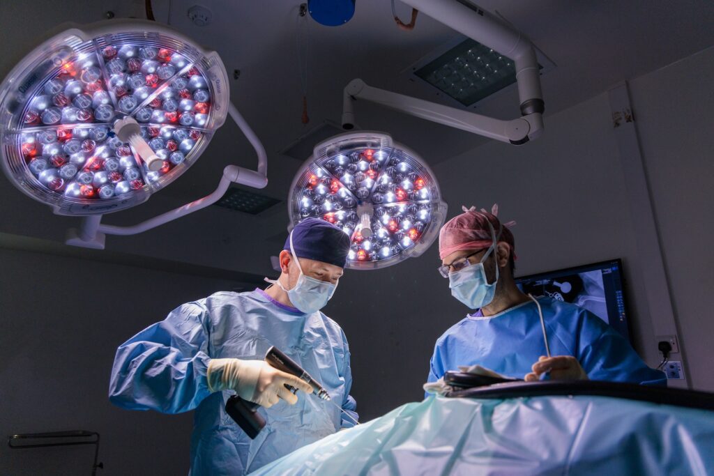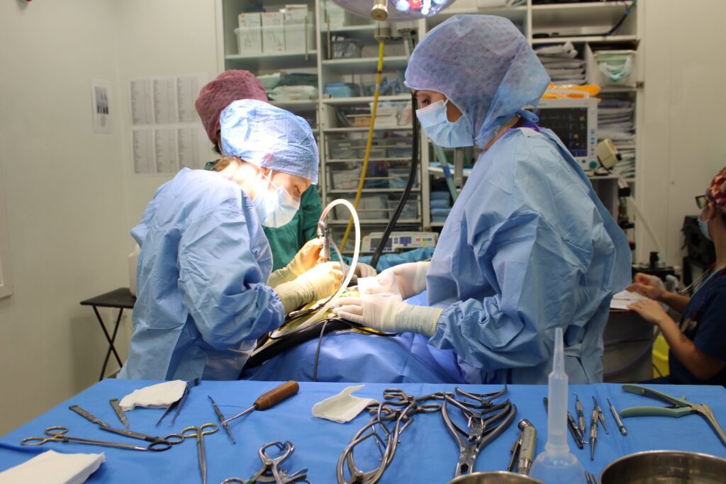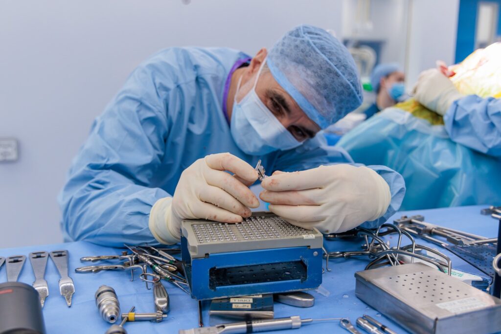
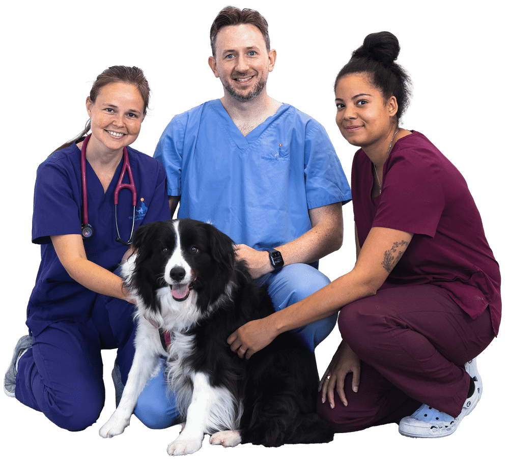
STATE-OF-THE-ART FACILITIES
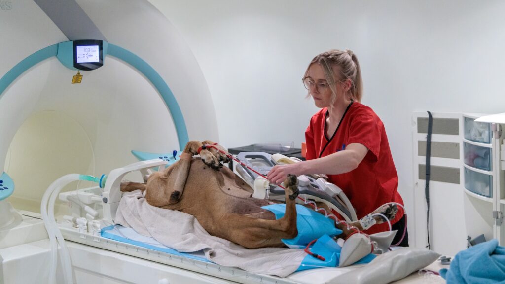
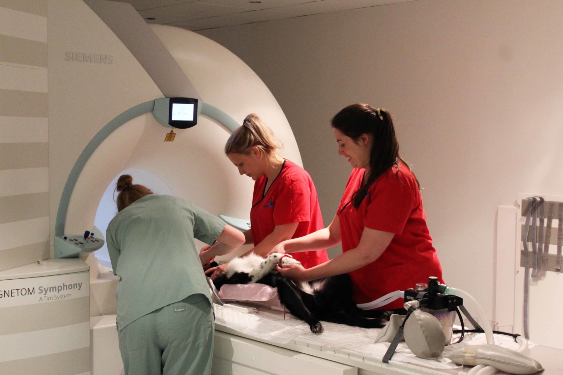
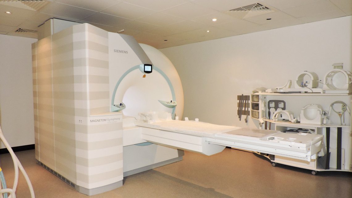
MRI machine
Our Siemens 1.5T MRI Symphony Tim system allows us to capture images of exceptional quality, ensuring efficient diagnoses and reduced scan times for our patients. This high field magnet system boasts 32 channels and the ability to combine up to 72 coil elements, meaning we are able to perform whole body scans without any repositioning and can move seamlessly from imaging one body area to another.
Since introducing the new MRI machine, we have already managed to reduce scanning time by 30%. As a result, patients have a reduced period of time under anaesthetic, which is particularly important in trauma cases.
What is MRI?
MRI works by using an extremely powerful magnet and a series of radio waves to stimulate water molecules in the body, which subsequently emit a signal. The advanced computer software is then able to collect all these signals and produce a map of where they all originate from. As water content differs between various anatomy the resulting image is able to demonstrate excellent contrast and differentiation between body structures, particularly soft tissue structures making it the modality of choice for the central nervous system.
Scans are tailored to the optimum parameters to demonstrate specific anatomies and pathologies in different contrasts so that a true assessment can be built up of what is happening inside the body. Our scanner is capable of using techniques including fat saturation, diffusion weighted imaging and three-dimensional acquisition to gain as much information as possible. Sometimes an intravenous injection of contrast media will be needed to further characterise areas of interest.
When is MRI used?
Common conditions that we would use MRI for include intervertebral disc disease, soft tissue and neurological tumours, epilepsy, syringomyelia, vestibular pathology, strokes or vascular events and ligament and tendon ruptures.
Due to the lengthier scan times,patients who undergo MRI will always require a GA. Unlike CT or X-ray, no ionising radiation is involved, but there are other safety considerations for these patients, namely the presence of metalwork or metallic foreign bodies as well as devices such as pacemakers and is important that we are aware of such objects before scanning.
Refer a patient today
Veterinary professionals – register to refer patients quickly and efficiently, and view your referral history.


CASE STUDIES
Patient stories
Our stories give an insight into some of our patients and their journey while having treatment at Fitzpatrick Referrals.
PATIENT STORY
Major Tom’s osteosarcoma
PATIENT STORY
Protected: Nell’s severe knee cartilage erosion
PATIENT STORY
Holly’s hydrotherapy rehabilitation after spinal surgery
PATIENT STORY
Shadow’s fractured canine tooth

PATIENT STORY
Leeroy’s elbow trauma injury
PATIENT STORY
Monty’s bilateral antebrachial growth deformity
PATIENT STORY
Ellie’s intervertebral disc extrusion (IVDE)
PATIENT STORY
Sunny’s rotating hinge total knee replacement
PATIENT STORY
Betsy’s amputation prosthesis
PATIENT STORY
Juno’s humeral condylar fracture

CAREERS
Join the team
We are always looking to strengthen our team with skilled, dedicated, passionate professionals.