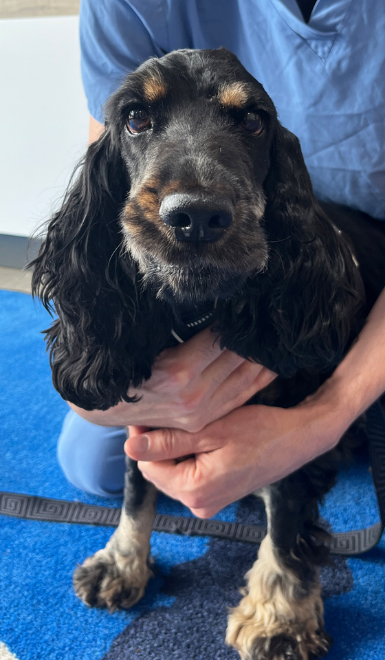
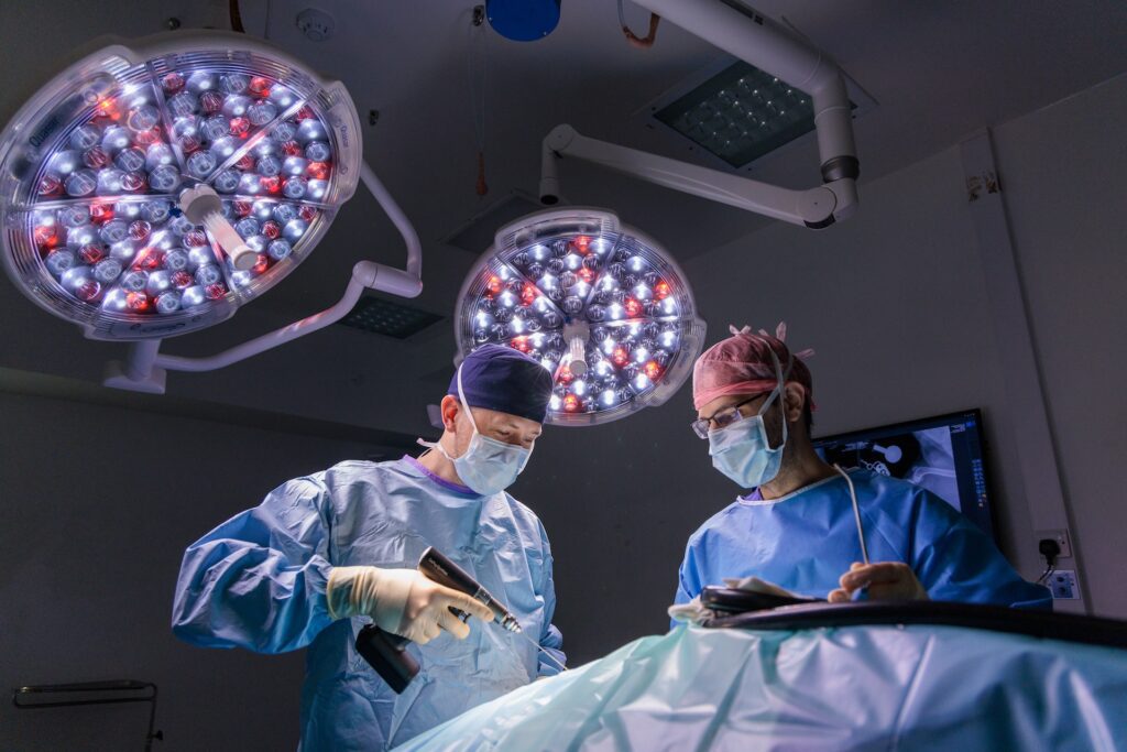
Under the care of RCVS Specialist in Small Animal Surgery Miguel Solano, Rosie had a CT scan and then arthroscopy of her right elbow.
CT scan
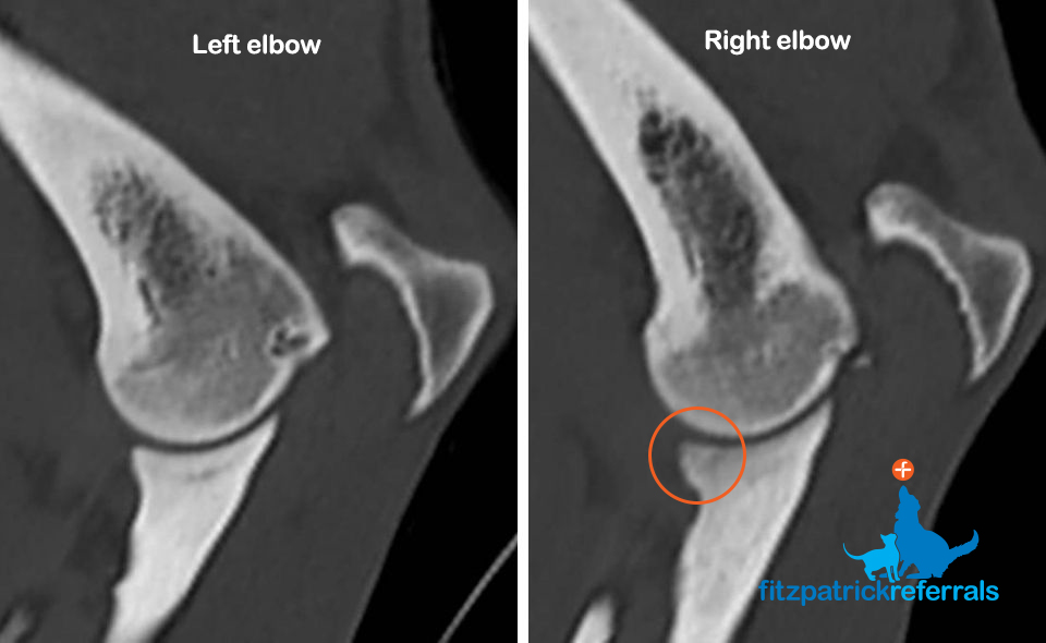
For clients: CT scan of the left and right elbows showing a darker tip of the ulna (medial coronoid) on her right confirming elbow dysplasia
Diagnosis: medial coronoid disease
Arthroscopy is a minimally invasive surgical procedure that allowed Miguel to look at the joint using a scope which acts as a magnified camera, to identify the cause of the lameness.
Diagnosed with medial coronoid disease, a form of developmental elbow disease, Miguel then performed a procedure called Subtotal Coronoid Ostectomy (SCO) to remove the diseased portion of her ulna.
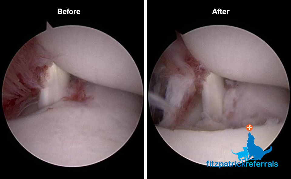
For clients: Keyhole surgery (arthroscopy) showing Rosie’s right elbow with signs of inflammation and some degree of cartilage wearing on the tip of the ulna (bottom) and on the humerus (top). Same elbow after removal of the diseased portion of the ulna (medial coronoid) to improve her discomfort.
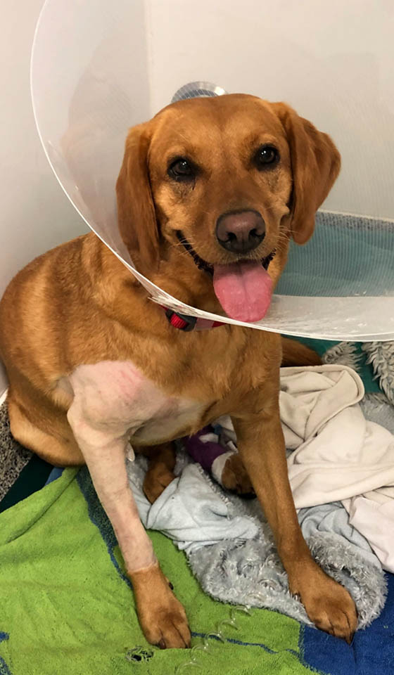
Here is the lovely Rosie pictured recovering in the wards after her surgery.
She headed home back to her family the same day as her procedure and will continue her recovery with six weeks’ rest and restricted exercise, followed by a rehabilitation programme of physiotherapy and hydrotherapy.
Read more about canine developmental elbow disease.
Services used in this story
Fitzpatrick Referrals is dedicated to the prevention and treatment of orthopaedic, neurological and dental diseases in small animals.